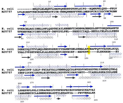
Figure 2.
Secondary structure alignment of E. coli TS and MJ0757. Sequence alignment (in single-letter code) generated by orf, based on predicted secondary structure. Observed secondary structure (blue), from x-ray coordinates used in Fig. 1, is shown above the E. coli sequence, with α-helices indicated by hatched boxes and β-strands by arrows. Secondary structure predicted by both gor (25) (blue) and phd (gray) (38) is shown below the MJ0757 sequence. The position of the catalytic cysteine is highlighted in yellow. Residues are numbered for convenience.

