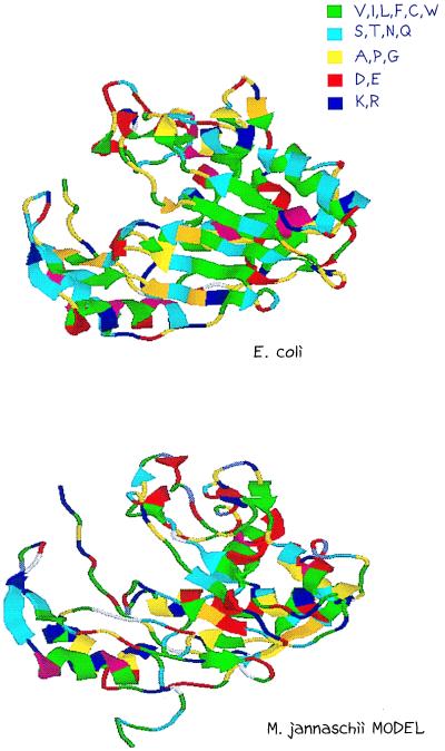
Figure 4.
Three-dimensional model of MJ0757. Using the program look 2.0, the MJ0757 sequence was threaded onto the E. coli structure. Side chain placement was refined in look 2.0 and further refined in x-plor (28). The model is visualized in rasmol (53) and color-coded by residue property: hydrophobic (V, I, L, M, F, W, and C), polar (S, T, N, and Q), special backbone (A, P, and G), positively charged (K and R), and negatively charged (D and E). The E. coli x-ray structure (Upper) and MJ0757 modeled structure (Lower) are shown separately for comparison.

