
Figure 3.
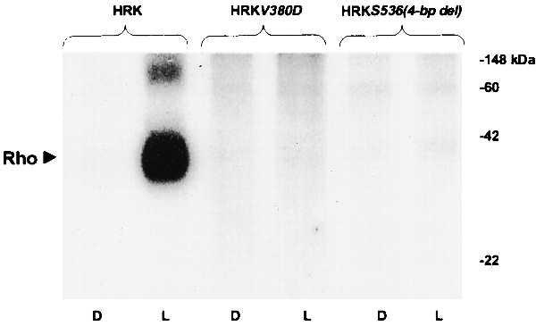
Rhodopsin phosphorylation by wild-type RK and the mutant forms HRKV380D and HRKS536(4-bp del). Autoradiogram of electrophoretically fractionated urea-washed rod outer segments incubated with supernatants from COS7 cells transfected with expression plasmids encoding wild-type HRK, HRKV380D, and HRKS536(4-bp del) in the presence of [γ-32P]ATP for 20 min in the dark (D) or the light (L). The enzyme assays were run at saturating substrate concentrations as detailed in Materials and Methods. The levels of RK immunoreactive protein in each assay are indicated by the arrowheads in Fig. 2B. The reactions were quenched by addition of SDS loading buffer at room temperature and an aliquot from each, containing 0.7 nmol of rhodopsin, was fractionated on 10% SDS-polyacrylamide gels before autoradiography. The positions of rhodopsin and molecular weight standards are marked.