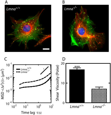
FIGURE 1.
Altered mechanical properties of the cytoplasm in MEFs lacking lamin A/C. (A and B) Immunofluorescence micrographs of the actin filament (green) and microtubule (red) networks in Lmna+/+ MEFs (A) and Lmna−/− MEFs (B), overlaid with fluorescence micrographs of ballistically injected 100-nm diameter nanoparticles (yellow). Cells were fixed and actin, microtubule, and nuclear DNA were stained using Alexa-488 phallodin, α-tubulin/Alexa568, and DAPI, respectively. Nanoparticles were enlarged for ease of visualization. Bar, 20 μm. (C) Average mean-squared displacements (MSD) of nanoparticles imbedded in the cytoplasm of Lmna+/+ MEFs (solid line) and Lmna−/− MEFs (dashed line) (n > 35 cells each). A higher value of MSD indicates larger movements of the particles, which indicates a softer cytoplasm. (D) Average shear viscosity of the cytoplasm of Lmna+/+ MEFs (left) and Lmna−/− MEFs (right) calculated from the MSDs (n > 35 cells each).

