FIGURE 5.
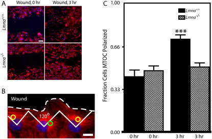
Impaired MTOC repositioning in MEFs lacking lamin A/C. (A) Lmna+/+ MEFs and Lmna−/− MEFs immediately after a wound and 3 h after the wound. Cells were fixed and microtubule and nuclear DNA were stained using α-tubulin/Alexa568 and DAPI, respectively. Bar, 100 μm. (B) Microtubule network organization in Lmna+/+ MEFs at the edge of the wound. Cells at the edge of the wound, which have their MTOC preferentially located within the front third facing the wound, are considered polarized (yellow circles); cells that have their MTOC located in the back two-thirds of the cell are considered nonpolarized (green circle). Bar, 20 μm. (C) Fractions of Lmna+/+ MEFs and Lmna−/− MEFs that had a polarized MTOC before the wound (0 h) and 3 h after wounding. For Lmna+/+ MEFs, n = 91 cells at 0 h and n = 175 cells at 3 h. For Lmna−/− MEFs, n = 183 cells at 0 h and n = 242 cells at 3 h.

