
Figure 1.
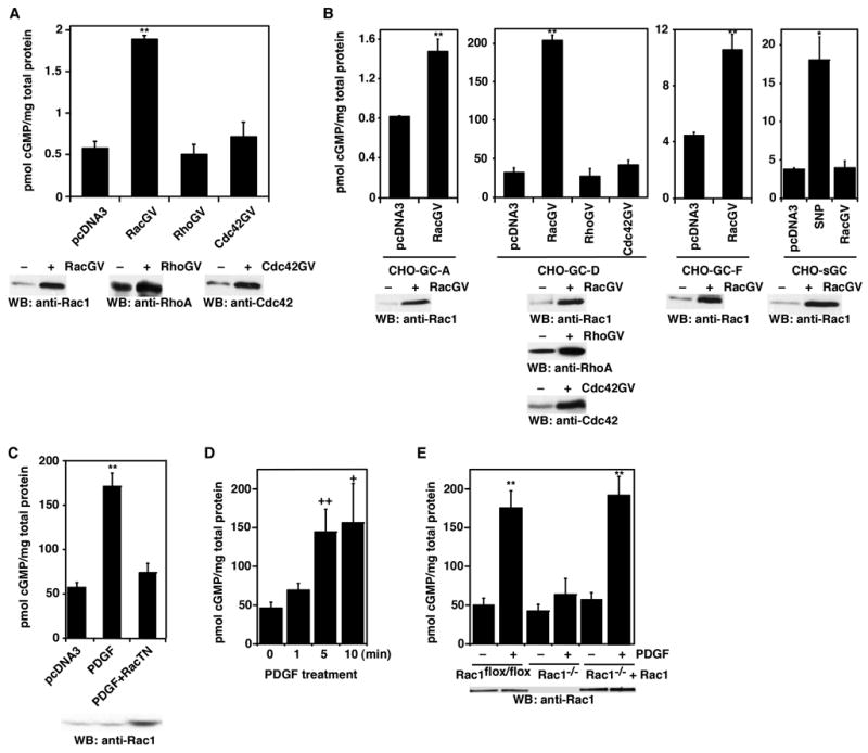
Rac1 stimulates GC activity in cells. A, Increase of GC-E activity by Rac1 in CHO-GC-E cells. Rac1(G12V), RhoA(G14V), CDC42(G12V), and the control vector pcDNA3 plasmid DNAs were transiently transfected into CHO cells that stably express GC-E. Cellular cGMP levels were determined. Bottom panels: Western blots show the expression of transfected Rac1(G12V), RhoA(G14V), and Cdc42(G12V) proteins, comparing to pcDNA3 vector-transfected cells. B, Effect of Rac1 on the activity of GC-A, GC-D, GC-F and soluble GC. Rac1(G12V) and the control vector pcDNA3 plasmid DNAs were transiently transfected into CHO cells stably expressing GC-A, GC-D, GC-F, or sGC (α1β1 isoform). Cellular cGMP levels were determined. Bottom panels: Western blots show the expression of transfected Rac1(G12V) proteins, comparing to pcDNA3 vector-transfected cells. C, Dominant-negative Rac1(T17N) inhibited the stimulation of GC-D by PDGF. Plasmid DNAs for Rac1(T17N) or pcDNA3 vector were transiently transfected into CHO-GC-D cells. Cellular cGMP levels were measured with or without PDGF stimulation (20 ng/ml for 5 min). D, Time course of cGMP accumulation after PDGF treatment. Data are shown as mean ± s.d. of three to five experiments. E, Rac1 deficiency blocked the stimulation of GC-D by PDGF. Rac1flox/flox cells, before or after infection of Cre-retrovirus, stably transfected with GC-D were treated with or without PDGF (20 ng/ml for 5 min). Rac1−/− +Rac1 cells were Rac1−/− cells infected with retroviruses containing wild-type Rac1. Cellular cGMP levels were measured. Error bars show mean ± s.d., **, P < 0.001; *, P < 0.005; ++, P < 0.01; +, P < 0.05 (Student’s t-test).