
Figure 5.
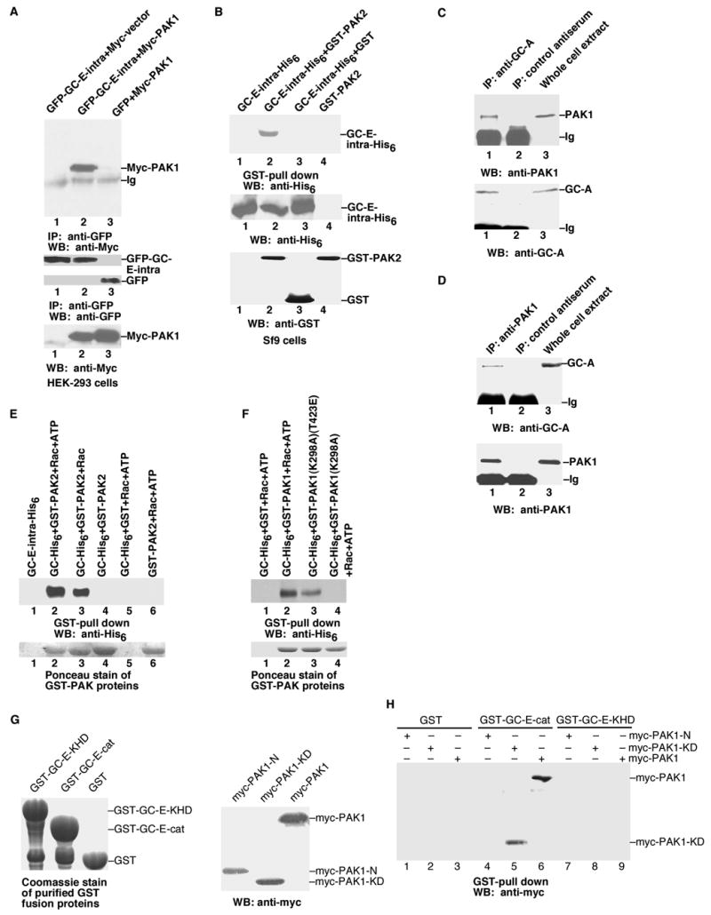
Direct interaction of PAK and GC-E. A, Co-immunoprecipitation of PAK1 and GC-E in HEK-293 cells. Bottom panels: anti-myc or anti-GFP antibodies were used to detect the protein expression of transfected genes in whole cell extracts. B, Co-immunoprecipitation of PAK2 and GC-E in Sf9 cells. C, PAK1 interacts with GC-A in vivo. Whole-cell lysates (MEF cells transfected with Rac1(G12V) to activate endogenous PAK) were subjected to immunoprecipitation with anti-GC-A antibody and immunoprecipitated proteins were analysed by immunoblotting with anti-PAK1 antibody. D, Anti-PAK1 antibody co-immunoprecipitated GC-A from MEF cells. Conditions were the same as in C. E and F, Interaction of purified PAK with purified GC-E in the presence of Rac-GTPγS. G Left panel: Coomassie staining of purified GST-GC-E-KHD (the kinase homology domain, residues 488 to 829), GST-GC-E-cat (the catalytic cyclase domain, residues 830 to 1059), or GST proteins used in the interaction assays. Right panel: Western blot with anti-myc antibody to show the expression of transfected myc-PAK1-N (residues 1 to 270), myc-PAK1-KD (residues 271 to 544), and myc-PAK1 in HEK-293 cells used for the interaction assays. H, Interaction of GC-E and PAK1. Purified GST, GST-GC-E-cat, or GST-GC-E-KHD proteins were mixed with lysates from HEK-293 cells transfected with myc-PAK1-N, myc-PAK1-KD, or myc-PAK1. Data are representative of three to four similar experiments.