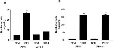
Figure 5.
IGF-I- and PDGF-stimulated cell migration in cells expressing full-length IAP and IAPc-s. Confluent cells were wounded then incubated ± IGF-I (100 ng/ml) (A) or PDGF (10 ng/ml) (B) for 48 h. The number of cells migrating across the wound edge in at least five preselected regions was counted. Each data point represents the mean ± SEM of three independent experiments. **p < 0.05 when migration of cells expressing IAPfl in the presence of IGF-I or PDGF is compared with incubation in SFM alone or when cells expressing IAPc-s stimulated with PDGF are compared with SFM alone.

