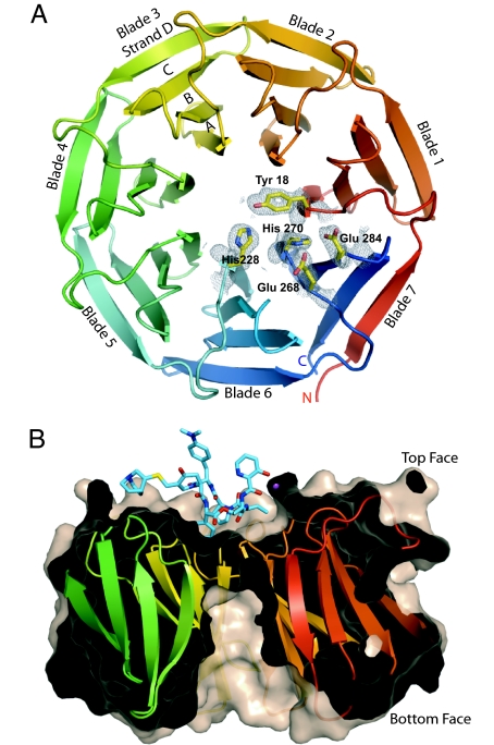
Fig. 1.
Structure of Vgb from S. aureus. (A) Ribbon diagram of Vgb viewed down the central axis. Five active-site residues are also shown together with their corresponding 2 Fo − Fc density map contoured at 1σ, as observed in the 1.65-Å structure. (B) View rotated 90° about the horizontal axis. The structure is sliced through the center to highlight the depression and the tunnel located on the top and bottom face of Vgb, respectively. Also shown is quinupristin, which binds in the depression.

