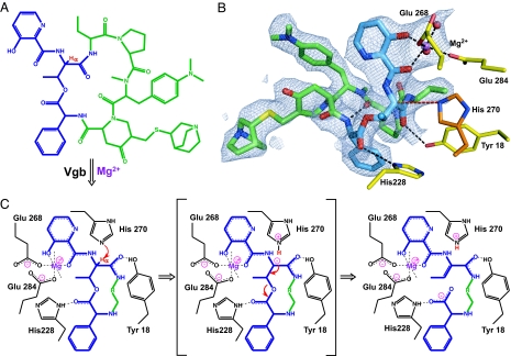
Fig. 2.
Proposed reaction mechanism for Vgb lyase activity. (A) Chemical structure of quinupristin. The 3-hydroxypicolinic acid, threonyl, and phenylglycyl moieties are colored blue, and the threonyl α-proton is shown in red. (B) Active site of Vgb. Quinupristin is shown using the same color scheme as in A, Mg2+ is shown in purple, active-site residues are displayed in yellow, with the modeled catalytic base His-270 displayed in dark orange, and water molecules are shown as red spheres. Also displayed is the 2Fo − Fc density map contoured at 1σ for quinupristin and Mg2+. (C) Schematic drawing of the proposed reaction mechanism. Note that only a part of quinupristin is shown, the remainder is represented by R.

