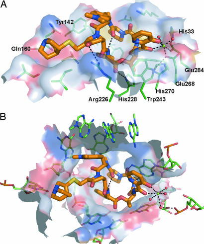
Fig. 3.
Comparison between quinupristin binding to Vgb and the ribosome. (A) Shown is the surface of Vgb within 5.5 Å of the bound streptogramin. The surface is colored according to the identity of the associated atoms: N, blue; O, red; C, white; S, yellow. Also shown are residues that contribute significantly to the surface, as well as Mg2+ and the coordinated water molecules. (B) Identical diagram for the 50S ribosomal subunit (Protein Data Bank ID code 1YJW). The view in both images is such that the orientation of the quinupristin matches in the two complexes (rmsd for all quinupristin ring atoms is 0.22 Å).

