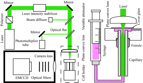
Fig. 1.
Schematic drawing of the apparatus developed for the flow detection of single molecules. (Right) A syringe pump was used to supply the solution of labeled protein into a capillary cell, which was made by fused silica with an inner diameter of 75 μm. (Left) A laser beam at 532 nm was passed through a polarizer and an intensity stabilizer and was focused to the sample exit of the capillary. The laser intensity was monitored by a photomultiplier tube. The florescence emitted from single molecules was collected by a camera lens, passed through four optical filters, and imaged on an EMCCD.

