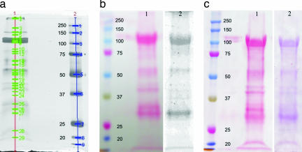
Fig. 3.
SDS/PAGE of the watery saliva of M. viciae and labeling of calcium-binding proteins with radioactive 45Ca2+ or by staining with ruthenium red on a Western blot nitrocellulose membrane. (a) Saliva concentrate of 1,000 aphids separated on a 10% gel (lane 1). Lane 2 presents marker proteins (kDa; Precision Plus Protein All Blue Standards; Bio-Rad, Hercules, CA). The silver-stained protein bands are marked and numbered by use of Quantity One 1-D Analysis Software (Bio-Rad). (b) Saliva collected from 41,000 aphids and separated on 10% SDS/PAGE and blotted on a nitrocellulose membrane. Lane 1 shows an overview of the separated proteins stained with Ponceau S, whereas the left lane presents marker proteins. In lane 2, after destaining the membrane, calcium-binding proteins are labeled with radioactive 45Ca2+, and radiolabeled proteins are detected by a PhosphorImager. (c) Lane 1 shows an overview of all separated proteins stained with Ponceau S, whereas the left lane presents marker proteins. In lane 2, after destaining the membrane, calcium-binding proteins are identified by ruthenium red staining.

