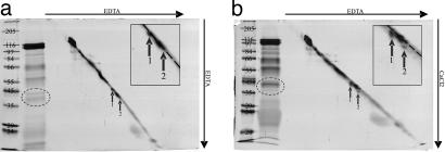
Fig. 4.
Two-dimensional SDS/PAGE of watery saliva of M. viciae in 10% separation gels. Saliva concentrate from 3,000 aphids is separated in two dimensions by common SDS/PAGE (no isoelectric focusing in the first dimension, as usual). The lane that contains saliva concentrate and diagonal marker is excised from first-dimension gel and placed perpendicularly to the running direction onto a second gel. Gel a contains EDTA in first and second dimension, whereas gel b contains EDTA in the first and CaCl2 in the second dimension. Proteins 43 kDa (1) and 40 kDa (2), indicated by arrows, shift downwards in the presence of Ca2+. In insets of the zoomed region of interest, the dotted line shows the position of the diagonal marker. Proteins are silver stained. The gel lane on the far left of each figure presents standard weight markers (Marker-Wide molecular weight range; Sigma–Aldrich; left lane, gel a, kDa) for the second-dimension separation. Next to the lane with the markers is a 1D lane of watery saliva proteins (1,000 aphids).

