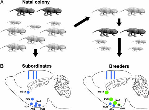
Fig. 1.
Social status affects reproductive brain nuclei in naked mole-rats. (A) All subjects initially were adult subordinates (light gray) within the natal colony, which also contains breeders (dark gray) and pups of various ages. One-half of the animals were randomly assigned to remain as colony subordinates (Left). The other one-half were removed from the colony and housed with a subordinate of the opposite sex (Right). These paired animals were defined as breeders when they had produced at least one litter. (B Left) Schematic of the sagittal plane of the brain of a subordinate naked mole-rat illustrating the approximate location of the brain regions examined. Hypothalamic and limbic nuclei are indicated by blue circles; rectangles indicate sites of cortical thickness measurements. (Right) The brain of an animal that has transitioned to breeding status. Green circles indicate regions that are either significantly larger (BSTp, PVN, and MeA) or have more cells (black dots, VMH) in breeders. Regions shaded blue did not differ between subordinates and breeders (SCN, ACo, and cortical thickness).

