Fig. 1.
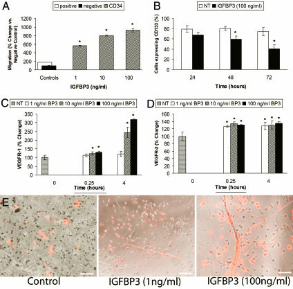
IGFBP3 modulates CD34+ cell behavior in vitro. (A) CD34+ cells (hatched bars) migrated in a dose-dependent manner toward IGFBP3. Migration is measured by relative fluorescent units (RFUs) compared to negative control. Negative control, medium alone arbitrarily set at 100 (filled bar); positive control, HPGM medium containing 20% serum (open bar). (B) CD34+ cells exposed to IGFBP3 (filled bars) for 48 and 72 h demonstrate reduced CD133 expression, supporting that IGFBP3 promotes their differentiation toward endothelial cells (P < 0.05 vs. control shown in open bars). (C) IGFBP3 exposure significantly increased VEGFR1 expression in CD34+ cells at the higher concentrations tested. ∗, P < 0.05 vs. control for 15 min and P < 0.001 for 4 h. (D) IGFBP3 increased the expression of VEGFR2 by 26.58% (∗, P < 0.001) at 15 min of exposure and by 27.5% (P < 0.001) at 4 h of exposure. (E) IGFBP3 increases EPC proliferation and tube formation compared to cells treated with control medium. IGFBP3 exposure resulted in a dose-dependent increase in tube formation. The cells have been exposed to fluorescently labeled acetylated LDL. The labeled cells appearing “red” in color represent EPCs that have differentiated into endothelial cells. (Magnification: ×100.) (Scale bars: Left and Center, 150 μm; Right, 100 μm.)

