Fig. 5.
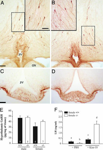
GnRH neurons in the hypothalamus of Kiss1tm1PTL-null mice and responses to kisspeptin injection. Photomicrographs of 50-μm-thick coronal sections showing GnRH immunoreactive neurons in the hypothalamus of wild-type (A and C) and mutant (B and D) mice. (A and B) GnRH-positive cell bodies in the preoptic region at the level of the organum vasculosum laminae terminalis. (Scale bar: 100 μm.) Frames in top corners are higher magnifications of respective dotted line squared areas. (Scale bar: 50 μm.) (C and D) GnRH-positive axonal projections and nerve terminals in the median eminence. (Scale bar: 50 μm.) 3V, third ventricle; ON, optic nerve. (E) GnRH content in hypothalami from both sexes. (F) Stimulation of LH release by kisspeptin-10 in Kiss1tm1PTL-null mice. Kiss1-null (−/−) or wild-type (+/+) female mice at diestrus were injected i.p. with vehicle (PBS) or 1 nmol of kisspeptin-10 in PBS and killed after 30 min, and serum was measured for LH. a, b, c, and d indicate values significantly different from each other (P < 0.05, n = 6 per group, one-way ANOVA followed by Student–Newman–Keuls test).

