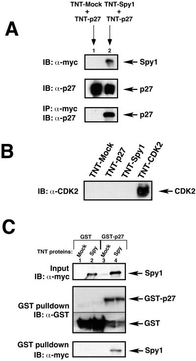
Figure 3.
Human p27 interacts with human Spy1 in vitro. (A) In vitro–translated p27 (TNT-p27) was incubated for 2 h with either in vitro–translated pCS3-MT empty vector (TNT-mock) or in vitro–translated myc-tagged Spy1 (TNT-Spy1). The top panel demonstrates the amount of input TNT-Spy1 protein (lane 2), the middle panel demonstrates the input TNT-p27 protein (lanes 1 and 2). Both samples were immunoprecipitated with α-myc sera, and proteins were separated by 10% SDS-PAGE and immunoblotted with p27 sera (bottom panel). (B) In vitro–translated pCS3-MT (TNT-mock), p27 (TNT-p27), myc-Spy1 (TNT-Spy1), or CDK2 (TNT-CDK2) were separated by 10% SDS-PAGE and immunoblotted with α-CDK2 sera as a control. (C) Purified GST-p27 or GST-control protein conjugated to GST beads were incubated in binding buffer with either in vitro–translated (TNT) mock or Spy1 protein (top panel), both of which were previously immunodepleted for any CDK2 protein as an additional control. A GST pull-down assay was performed. Proteins were separated by 12.5% SDS-PAGE and immunoblotted with α-myc sera to detect immunoprecipitation of GST proteins with myc-tagged proteins (bottom panel). α-GST immunoblot is presented to demonstrate the GST proteins present in the GST pull-down experiment. Different exposures of the same immunoblot are presented in the middle panel because of the high amount of protein in the GST-control lanes.

