
Fig. 2. Multinucleation caused by GEF-H1 perturbation.
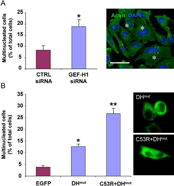
(A) Multinucleated cells were found at 48 h after transfection with GEF-H1 siRNA. Values shown are the means from four independent experiments in which over 1000 cells were counted per experiment; error bars indicate SEM. For statistical analysis, all data were evaluated by two-tailed Student’s t-test. Values significantly different from controls (p≤ 0.01) are marked with an asterisk. Cells harboring more than 1 nucleus are indicated by an asterisk in the micrograph. Scale bar represents 20 μm.
(B) Overexpression of GEF-H1 inhibitory mutants induced multinucleation. The number of the multinucleated cells as a percentage of the total cell population expressing the EGFP-tagged constructs was quantified in HeLa cells 48 h after transfection. A minimum of 100 cells from each of three independent experiments was scored for each construct. Values significantly different from EGFP-expressing cells are marked with one (p≤0.01) or two asterisks (p ≤0.001). Micrographs on the right illustrate expression patterns of the different constructs.