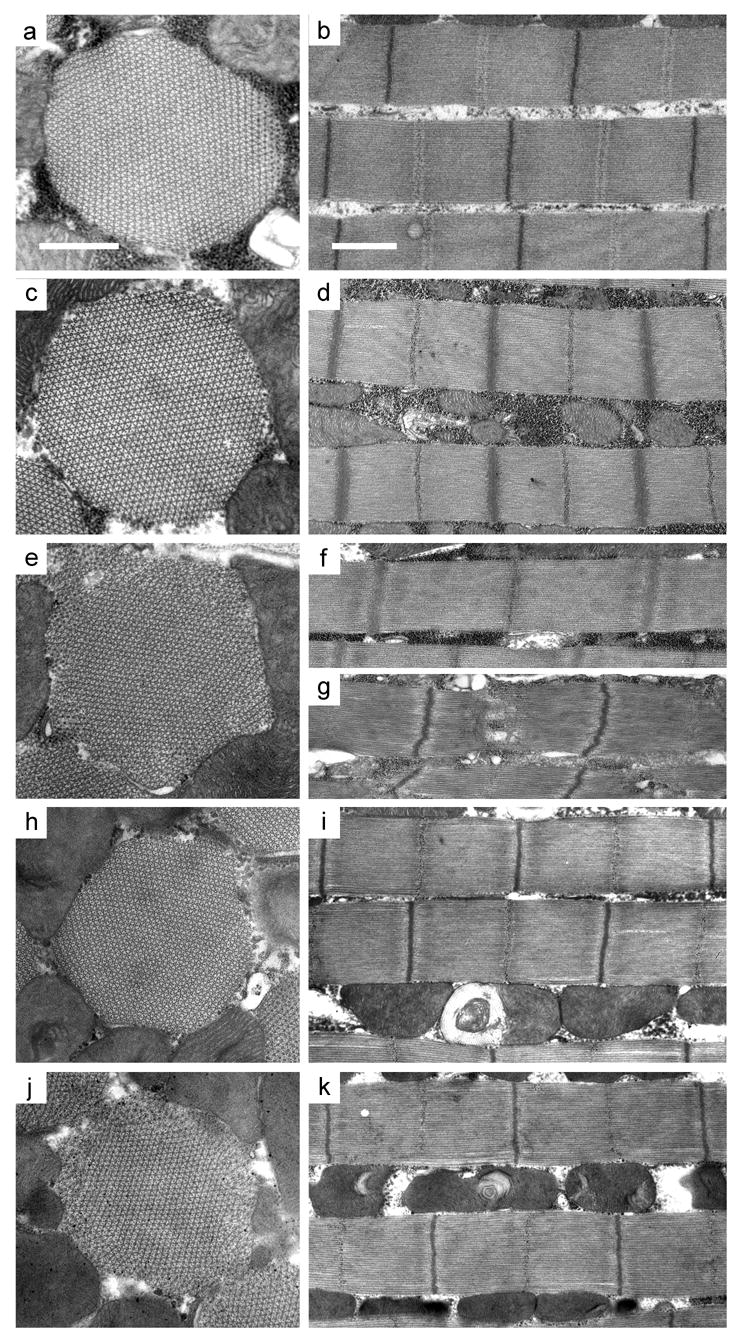
Figure 3.

Electron micrographs of myofibrils from 2-day old wild-type (yw), PwMhc2 control and PwMhc2-15b hinge-switch IFM. Transverse (left panels) and longitudinal (right panels) sections are depicted. (a) IFM of yw (wild-type) has regular hexagonal thick and thin filament arrays and (b) intact sarcomeres with granule-containing M-lines and electron-dense Z-lines. (c, d) PwMhc2 myofibrils have a nearly wild-type structure; (c) 90–95% of myofibrils have intact double hexagonal packing of thick and thin filaments throughout and (d) normal M-line and Z-disk structure. There is some variation in sarcomere length in both yw and PwMhc2 lines, but the mean sarcomere lengths do not differ statistically (2.94 ± 0.02 μm for yw, 2.87 ± 0.02 μm for PwMhc2, mean ± SEM). Myofibrils in 15b-3 (e, f), −47 (h, i), and −108 (j, k) have varying degrees of minor disruption at 2 days post-eclosion, including peripheral filament packing defects. Some hinge-switch sarcomeres (f) are obviously longer than wild type (b) or control (d). Occasional severe disruption, particularly in the M-line region, is observed in the hinge-switch lines (g, line 15b-3). Transverse images, bar = 0.5 μm. Longitudinal images, bar = 1.0 μm.
