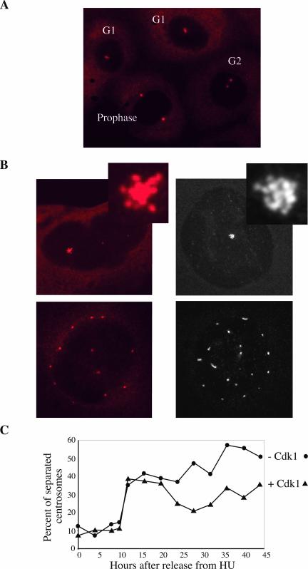
Figure 4.
Centrosome segregation timing is conserved in endoreplicating cells. (A) Normally growing cells stained with a γ-tubulin antibody with clustered (G1), separated (G2), and fully segregated (prophase) centrosomes. (B) Endoreplicating cells stained with γ-tubulin antibodies were imaged either by wide field (left) or by confocal microscopy (right). The confocal image is a projection of the series of z-stacks spanning the entire depth of the cell to show centrosomes at all focal planes. The centrosomes in cells were either clustered (top) or segregated (bottom). (C) Cells cultured without IPTG for 7 d (–Cdk1) and control cells (+ Cdk1) were arrested with HU and released into fresh medium. Cells were fixed at the indicated times and stained with a γ-tubulin antibody. Two hundred cells of each time point were photographed to score the proportion of cells with separated centrosomes.

