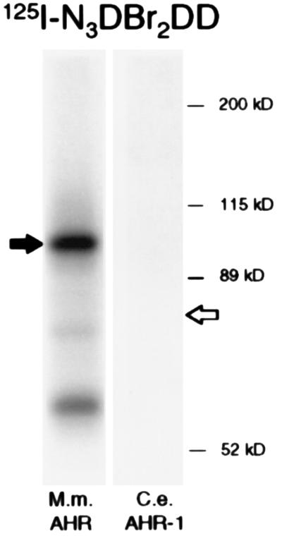
Figure 4.
Photoaffinity labeling of AHR-1 and AHR with a dioxin analog. C. elegans AHR-1 and mammalian AHR were expressed in rabbit reticulocyte lysates. The proteins were incubated with 125I-N3Br2DD, photoaffinity labeled, separated by SDS/PAGE, and visualized by autoradiography. The sizes of murine AHR and C. elegans AHR-1 are indicated by filled and open arrows, respectively. No labeling of AHR-1 could be detected.

