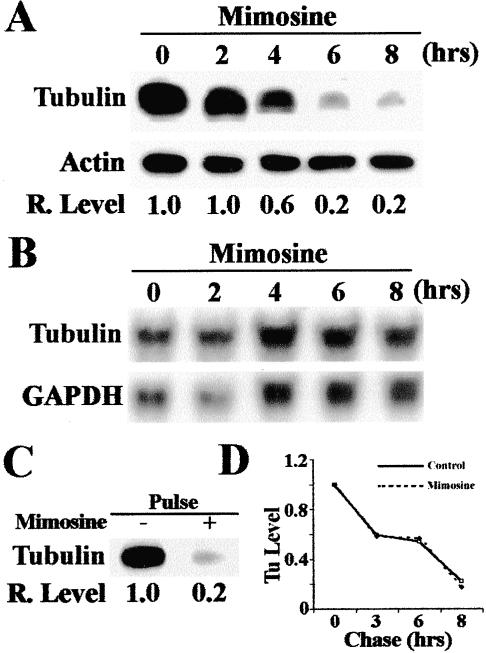
Figure 5.
Effect of mimosine on the expression of tyrosinated α-tubulin. (A) Time course of the effect of mimosine on the expression of tyrosinated α-tubulin. HeLa cells were treated with 600 μM mimosine for 0–8 h and then lysed for determination of the expression of tyrosinated α-tubulin by Western blot. Actin was used as loading control. (B) Northern blot analysis of mRNA level of α-tubulin after mimosine treatment. HeLa cells were treated with 600 μM mimosine and total RNAs were prepared for Northern blot analysis of α-tubulin. GAPDH was used as a loading control. (C) Effect of mimosine on the synthesis of tyrosinated α-tubulin. HeLa cells were treated with or without 600 μM mimosine for 6 h followed by a 2-h pulse labeling with [35S]methionine. Labeled tyrosinated α-tubulin was then immunoprecipitated for SDS-PAGE and autoradiography analysis as described in MATERIALS AND METHODS. (D) Effect of mimosine on the degradation of tyrosinated α-tubulin. HeLa cells were first pulse-labeled with [35S]methionine for 2 h followed by a chase in DMEM supplemented with 100 μg/ml unlabeled methionine in the presence or absence of 600 μM mimosine. Tyrosinated α-tubulin was then immunoprecipitated for SDS-PAGE and autoradiography followed by measurement of tyrosinated α-tubulin level with a Scion Image software.

