Figure 4.
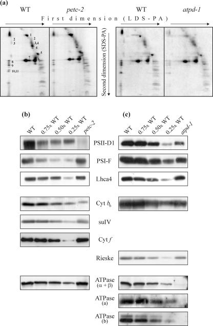
Protein composition of thylakoid membranes. a, Thylakoid membranes corresponding to 30 μg of Chl from WT, petc-2, and atpd-1 mutant plants were fractionated first by electrophoresis on a non-denaturing lithium dodecyl sulfate-polyacrylamide (PA) gel and then on a denaturing SDS-PA gel. Positions of WT thylakoid proteins previously identified by western analyses with appropriate antibodies are indicated by numbers to the right of the corresponding spots: 1, α- and β-subunits of the ATPase complex; 2, D1-D2 dimers; 3, CP47; 4, CP43; 5, oxygen-evolving complex (OEC); 6, LHCII monomer; 7, LHCII trimer; 8, PSI-D; 9, PSI-F; 10, PSI-C; and 11, PSI-H. The alterations observed in the mutants are quantified in Table I. Samples of thylakoid membranes equivalent to 5 μg of Chl from WT, and petc-2 (b) and atpd-1 (c) plants were fractionated by denaturing PAGE. Decreasing amounts of WT thylakoid membranes (3.75, 2.5, and 1.25 μg of Chl) were loaded in the lanes marked 0.75×, 0.5×, and 0.25× WT. Replicate filters were probed with antibodies raised against the D1 protein of the PSII reaction center, the F subunit of PSI, the Lhca4 protein, the cyt b6/f subunits cyt b6, cyt f and suIV, and the α-, β-, a-, and b-subunits of the cpATPase.

