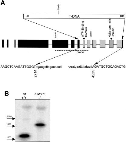
Figure 1.
Structure of T-DNA insertion in At-MSH2. A, Sequences of PCR products generated with gene-specific and T-DNA-specific primers were used to deduce the structure of the disrupted AtMSH2 in the line SALK_002708. A single insertion of pROK2 T-DNA at positions 2,714 and 4,225 caused deletion of exons 8 to 12 and portions of exons 7 and 13 (gray boxes) in this line. Sequences of junction regions are below. Capital letters indicate AtMSH2 and lowercase letters indicate the insertion, beginning 2 bp downstream of the left border at the exon 7 junction and preceded by approximately 150 bp of rearranged sequence following the right border at the exon 13 junction. B, DNA blot of EcoRV-digested wild-type and T-DNA insertion homozygotes probed with a radiolabeled At-MSH2 fragment (dashed line).

