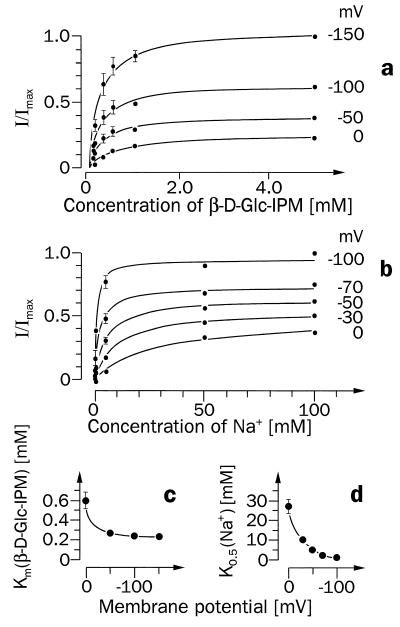
Figure 5.
Potential dependence of SAAT1-mediated currents induced by different concentrations of β-d-Glc-IPM in the presence of Na+ (a and c) or by different Na+ concentrations in the presence of β-d-Glc-IPM (b and d). Ten nanograms of SAAT1-cRNA was injected into Xenopus oocytes, and the oocytes were incubated for 5 days. (a) The currents are shown that were measured when oocytes clamped at different membrane potentials were superfused with different concentrations of β-d-Glc-IPM in the presence of 100 mM Na+. The measurements from three oocytes were normalized to their maximal currents at −150 mV. The Michaelis–Menten equation was fitted to the data obtained at the respective membrane potential, and the Km values were plotted against the different membrane potentials (c). The currents measured after superfusion of the oocytes with 1 mM β-d-Glc-IPM in the presence of different concentrations of Na+ are shown in b. Here measurements from three oocytes were normalized to their current in the presence of 100 mM Na+ at −100 mV. Means and SEM values are presented, and the Michaelis–Menten equation was fitted to the currents obtained at each membrane potential. The K0.5 values for Na+ activation at different membrane potentials are presented in d.

