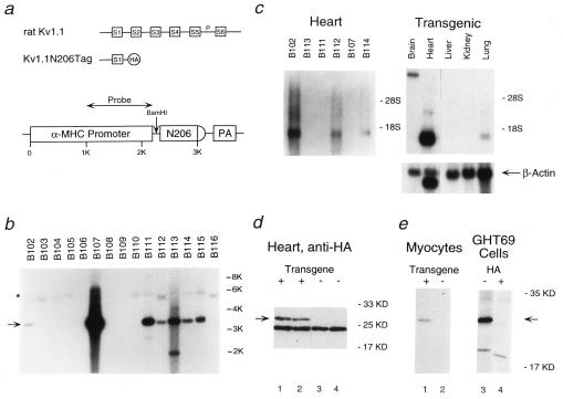
Figure 1.
Expression of the N-terminal fragment of rat Kv1.1 in the hearts of transgenic mice. (a) Schematic representation of rat brain potassium channel Kv1.1 (Top), the N-terminal fragment Kv1.1N206Tag (Middle), and the transgenic construct (Bottom). HA represents the HA epitope added to the N-terminal fragment of Kv1.1. The α-myosin heavy chain contained ≈1 KB of the promoter followed by exon 1, the first intron, and part of exon 2 (5′ untranslated). N206 represents the N-terminal fragment Kv1.1N206Tag and PA represents the polyadenylation signal. (b) Genomic Southern blot of tail DNA isolated from 16 Fo mice, digested with BamHI, and probed with the fragment of the α-myosin heavy chain denoted in a. The native mouse α-myosin heavy chain gene (∗) and the transgene (arrow) are indicated. Note that each of the seven transgenic founders have multiple copies of the transgene. (c) Northern blot analysis of 2 μg of poly(A)+ RNA (PolyATract, Promega) from the hearts of F1 mice from the lines indicated (Left) and of 15 μg of total RNA (RNEasy, Qiagen) from the indicated organs of an F1 mouse of the B102 line (Right), probed with the N-terminal fragment of Kv1.1. Relative loading of RNA was confirmed by reprobing with a 700-bp fragment of the β-actin gene. (d) Western blot analysis of the steady-state levels of Kv1.1N206Tag polypeptide present in the membrane fraction of LQT hearts (lanes 1–2) and control hearts (lanes 3–4) using a rabbit polyclonal antibody directed against the HA epitope (Santa Cruz Biotechnology). The arrow indicates the position of Kv1.1N206Tag. (e) Immunoprecipitation analysis of Kv1.1N206Tag expressed in primary culture of adult mouse rod shape cardiocytes derived from two LQT mice (lane 1), control cardiocytes (lane 2), and GH3 cells stably transfected with Kv1.1N206Tag (GHT69) (lane 3). In lane 4, the immunoprecipitation was blocked with 90 nmols of HA peptide. Total cell lysates were immunoprecipitated with anti-HA antibody (12CA5). The pellets were analyzed on SDS/PAGE. The arrow indicates the position of Kv1.1N206Tag.

