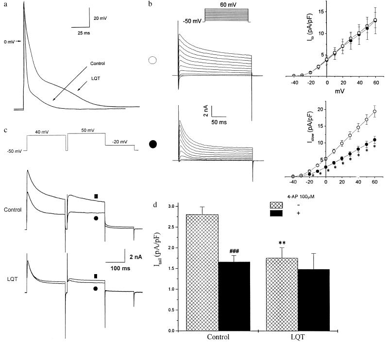
Figure 3.
Characterization of AP and the outward potassium currents in LQT cardiocytes. (a) Transmembrane AP recorded from a representative control and a LQT mouse cardiocyte. APs were elicited by suprathreshold current injected through the electrode at 0.5 Hz. For better superimposition, stimulatory artifacts resulting from current injection were minimized off-line. (b) Voltage-dependent K+ current traces and current-voltage relationships in control (∗; n = 24) and LQT mouse ventricular myocytes (•; n = 16). Traces from representative cells are shown (Left). Current density was obtained by normalizing the current amplitude to cell capacitance. The currents were elicited by 10 mV step depolarizations between −40 to +60 mV from −50 mV holding potential. Ito was defined as the difference between the peak outward current and the current level at the end of the 250-ms pulse. The current remaining after 250 ms was significantly lower in LQT myocytes (P < 0.05). (c) Effects of 50 μM 4-AP on the outward currents of a representative ventricular myocyte derived from either a control or a matched LQT mouse. The voltage protocol is shown (Top). ▪ and • indicate the current traces before and after application of 4-AP respectively. (d) Summary of the effects of 100 μM 4-AP on Itail of control and LQT myocytes. Itail was defined as the difference between the peak and the current at the end of −20-mV pulse. The density of Itail was significantly larger in control cardiocytes compared with LQT (∗∗, P < 0.01). 4-AP abolished over 40% of Itail in control cardiocytes (###, P < 0.001), but had no significant effect in LQT cardiocytes.

