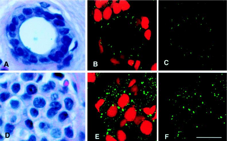
Figure 1.
Centrosome number and size in normal breast duct epithelia (A–C) and adenocarcinoma cells (D–F). Hematoxylin and eosin-stained paraffin sections of a normal human breast duct (A) and a breast adenocarcinoma (D). (B) Confocal image stack of a normal breast duct stained for centrioles with anticentrin mAb 20H5 (fluorescein isothiocyanate secondary antibody) and for nuclear DNA with propidium iodide. Approximately 20 pairs of centrioles are located apical to the nuclei of epithelial cells that line this normal duct. (C) Binary processed image showing the volume of centrin labeling for the same normal epithelial image stack shown in B, from which a portion of the data in Table 1 was derived. (E) Confocal image stack of a breast adenocarcinoma stained as above. Many large centrin-staining spots mark the location of abnormal centrosomes in the tumor tissue. (F) Binary processed image showing the volume of centrin labeling for the same tumor image stack shown in E, from which a portion of the data in Table 1 was derived. (Bar = 20 μm.)

