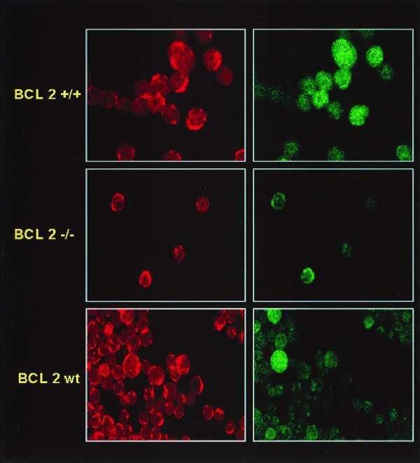
Figure 4.

Localization of GSH in mouse thymocytes expressing various levels of Bcl-2 as analyzed by confocal microscopy. Isolated thymocytes from mice transgenic for Bcl-2 (top), Bcl-2 knockout mice (middle), or control littermates (bottom) were stained with CMFDA and MTX and subjected to confocal microscopy. Mitochondria are highlighted by MTX localization in the left panels, whereas GSH localization is shown in the right panels. Of interest is the heterogeneous GSH staining between cells in the control mice suggesting high Bcl-2 levels in some thymocytes and low Bcl-2 levels in others.
