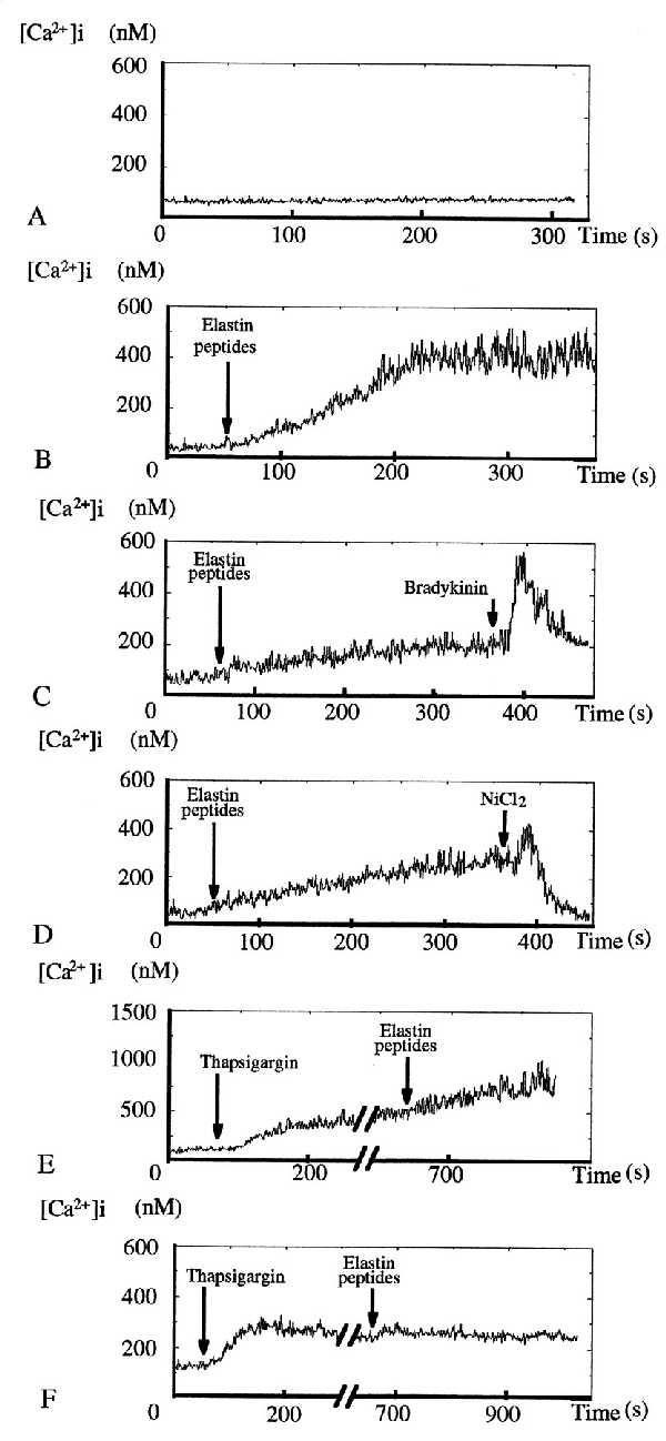
Figure 3.

Action of EP on suspended HUVEC [Ca2+]i. (A) Time-control tracing (n = 2). (B) Addition of 10−2 mg⋅ml−1 EP induced a slow but strong increase in [Ca2+]i, starting from a steady low [Ca2+]i before reaching a new high level steady state after 200 s (n = 10). [Ca2+]i increase amplitudes varied with the experiment and were from 1.5-fold up to 7-fold. (C) Addition of 1 μM bradykinin induced an immediate strong but short-lasting [Ca2+]i rise (n = 5). (D) Addition of 1 mM NiCl2 totally abolished EP-induced [Ca2+]i increase (n = 4). (E) Ten minute incubation in PSS ([Ca2+]o = 2.4 mM) with 1 μM thapsigargin had no effect on EP-induced [Ca2+]i increase pattern (n = 6). (F) EP became unable to induce a [Ca2+]i increase when the same experiments (n = 5) were performed in low calcium PSS ([Ca2+]o ≈ 10−7 M).
