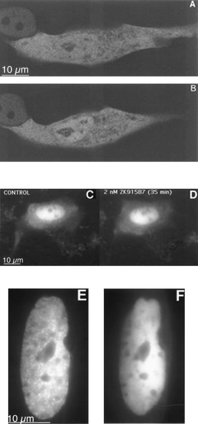
Figure 6.
Subcellular localization of the GFP-MR in the presence of MR antagonists. Confocal images of two cells before (A) and after (B) incubation with 1 μM spironolactone for 45 min at 37°C. C and D are fluorescence microscopic images of a cell before (C) and after (D) incubation with 2 nM ZK91587 for 35 min. In all three cells nuclear translocation of GFP-MR was significantly less than that with aldosterone. (E) Cells were perfused with 1 nM aldosterone for 30 min; note prominent nuclear clusters. After 30 min, the perfusion medium was changed to 1 nM aldosterone plus 100 nM spironolactone, and the image on F was captured 30 min later. Note that spironolactone treatment disrupted the nuclear clusters.

