Figure 1.
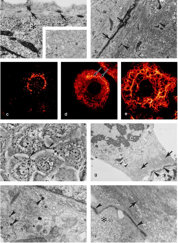
Expression of a cardiac-specific phenotype at multiple passages in the HL-1 cardiomyocyte cell line. (a) A 10th-passage HL-1 cell demonstrating subsarcolemmal Z densities (arrows) typical of normal myofibrillogenesis in cultured and in vivo cardiomyocytes, glycogen (∗). (×12,500.) (Inset) High-magnification view demonstrating typical cardiac-specific myofibril banding. (×20,000.) (b) Two 10th-passage HL-1 cells containing myofibrils at various stages of sarcomerogenesis (arrowheads) are attached via an immature intercalated disc (arrows). (×9,670.) (c) Immunofluorescent localization of ANF expression in a passage 20 HL-1 cell. (d) Myosin is localized to scattered filaments in the cytoplasm and to thin reorganizing myofibrils located along the peripheral cytoplasm (arrows) in a passage 20 HL-1 cell. (e) In a passage 20 HL-1 cell desmin is expressed as reticulated cytoplasmic rings of intermediate filaments extending into lamellapodia. (c–e) False color, indirect immunofluorescence confocal laser scanning microscopy images. (×1,650.) (f) Passage 34 HL-1 cells demonstrating centrally located mononucleation and intercellular junctional contacts (arrowheads); phase-contrast, ×700. (g) At passage 34, dividing HL-1 cells behave like typical mitotic cardiomyocytes in that they retain peripheral myofibrils (arrows), contain atrial granules (small arrowheads,) and are anchored to adjacent cardiomyocytes by intercalated discs (arrowheads). C, chromosomes. (×8,000.) (h) At passage 86, an active Golgi and atrial-specific granules are present (arrowheads). (×25,000.) (i) A passage 86 HL-1 cell containing organized myofibrils (arrow), an intercalated disc (arrowheads), and areas occupied by free ribosomes (∗). (×15,000.) (a, b, and g–i) Transmission electron micrographs.

