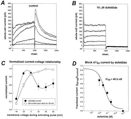
Figure 3.
Characteristics of IKr-like current recorded from HL-1 cells. (A) Whole-cell current traces recorded from an HL-1 cell. Voltage pulses (1-sec duration) were applied from a holding potential of −50 mV to test potentials between −40 mV to +40 mV in 20-mV increments. The membrane potential was returned to −50 mV after each depolarizing pulse to better resolve outward tail currents. (B) Whole-cell currents in the presence of 10 μM of dofetilide (same cell as in A). Dofetilide abolished the time-dependent component of the activating current and the deactivating tail currents. Complete blockade of the time-dependent component of the activating outward current revealed a residual time-independent outward current in most HL-1 cells. (C) Current-voltage relationship for the time-dependent activating outward current and the deactivating tail current. Plotted are the normalized magnitudes of whole-cell currents recorded at the end of each 1-sec depolarizing pulse (•) and tail currents (○) versus membrane potential. Currents were normalized as a fraction of peak current magnitude. Values are means ± SEM (n = 10 cells). (D) Concentration-response relationship for block of IKr tail current by dofetilide. IC50 was 46.9 nM (n = 4).

