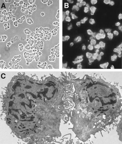
Figure 1.
Purity and appearance of type I cells. Cytocentrifuged cell preparation with (A) phase contrast and (B) immunofluorescence with a mAb specific for type I cells (C). Electron micrographs of isolated type I cells showing very thin cytoplasmic extensions typical of this cell type. Magnifications: A, ×280; B, ×300; C, ×4,150.

