
Figure 4.
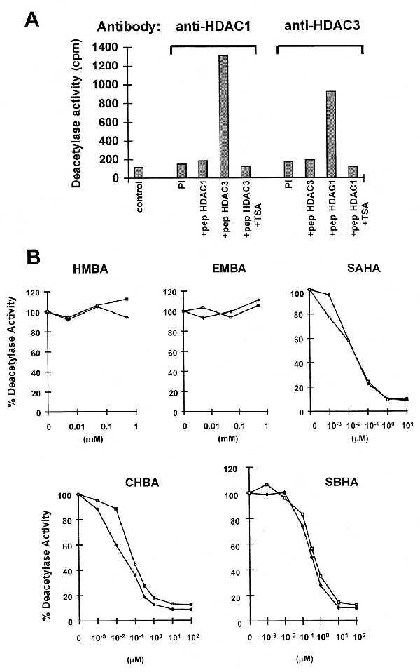
Inhibition of HDAC1 and HDAC3 by HPCs. (A) Cellular extracts from Jurkat cells (5 × 10⁷ cells) were subjected to immunoprecipitation with a polyclonal antiserum against HDAC1 and HDAC3 in the presence of a 100-fold excess of HDAC3 peptide (residues 413–428) or HDAC1 peptide (residues 467–482). As an additional control, the corresponding preimmune serum was used. Immunoprecipitated complexes were tested for HDAC activity by measuring the release of [3H]acetate from an acetylated amino-terminal H4 peptide (in cpm), in the absence or presence of 400 nM TSA. (B) Cellular extracts from Jurkat cells were subjected to immunoprecipitation with polyclonal antiserum against HDAC1 (♦) or HDAC3 (□) and tested for HDAC activity in the absence or the presence of increasing concentration of the following inhibitors: HMBA, 0.005, 0.05, and 0.5 mM; EMBA, 0.005, 0.05, and 0.5 mM; SAHA, 0.001, 0.01, 0.1, 1, and 10 μM; CBHA or SBHA, 0.001, 0.01, 0.1, 0.33, 1, 10, and 100 μM.