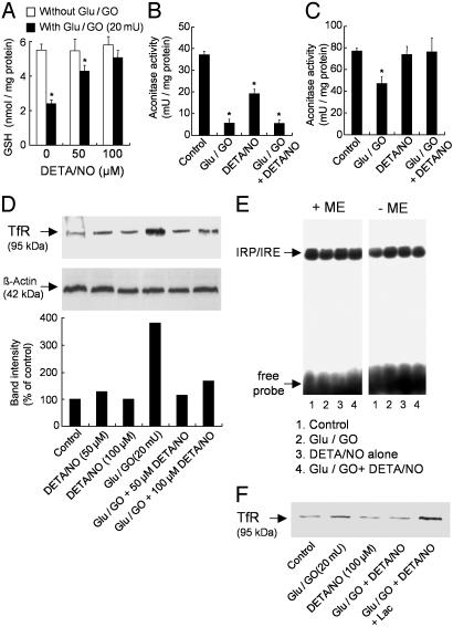
Fig. 2.
Effect of •NO on H2O2-induced changes in GSH, aconitase activity, TfR, and IRP-1 levels in BAECs. (A) BAECs were treated with glucose (Glu)/GO (20 milliunits) for 4 h with and without DETA/NO (50 and 100 μM), and GSH levels were measured. (B and C) Cells were treated with glucose/GO (20 milliunits) for 4 h with and without DETA/NO (100 μM), and aconitase activity was measured in the cytosolic (B) and mitochondrial (C) fractions. (D) BAECs were treated with glucose/GO for 4 h with and without DETA/NO, and TfR protein expression was measured by using 20 μg of protein of the 12,000 × g supernatant. (E) BAECs were treated as described for B, and cytoplasmic extracts were analyzed by gel-shift assay with and without 2-mercaptoethanol (2-ME) to measure IRP/IRE binding. (F) Cells were treated as described for D by using 12 μg of protein of the 12,000 × g supernatant except that the effect of lactacystin (10 μM) on TfR protein expression in glucose/GO-treated cells with and without DETA/NO was determined. *, P < 0.05 vs. control.

