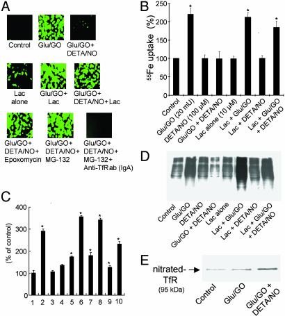
Fig. 3.
Effect of proteasomal inhibitors on DCFH oxidation, iron uptake, and apoptosis in BAECs treated with H2O2 and •NO. (A) BAECs were treated with glucose (Glu)/GO with or without DETA/NO (100 μM), proteasome inhibitors, lactacystin (10 μM), epoxomycin (2 μM) and MG-132 (10 μM), and anti-TfR antibody (IgA class, 42/6) (12 μg/ml). Cells were pretreated with the proteasome inhibitors and DETA/NO for 2 h before treatment with H2O2 for 4 h. Cells then were washed in DPBS and incubated with DCFH-DA (10 μM) for 20 min. Green fluorescence due to DCF was monitored with time. (B) 55Fe uptake into cells was monitored under the same conditions as described for A and C. Caspase-3 activity was measured under the same conditions described for A except that cells were treated with H2O2 for 8 h. 1, control; 2, glucose/GO; 3, DETA/NO alone; 4, glucose/GO + DETA/NO; 5, Lac alone; 6, Lac + glucose/GO; 7, Lac + DETA/NO; 8, Lac + glucose/GO + DETA/NO; 9, glucose/GO + AcOM-PYRRO/NO; 10, Lac + glucose/GO + AcOM-PYRRO/NO. (D) Cells were treated as described for B, and 5 μg of the derivatized total protein was resolved on an SDS/8% PAGE gel and probed with a monoclonal anti-2,4-dinitrophenol antibody to measure protein carbonyl levels by Western blot analysis. (E) BAECs were treated as described for A, and the cell lysate (100 μg of protein) was immunoprecipitated with 6 μg of anti-TfR antibody, resolved on an SDS/8% PAGE, and probed for nitrated TfR with anti-nitrotyrosine antibody by Western blot analysis. *, P < 0.05 vs. control.

