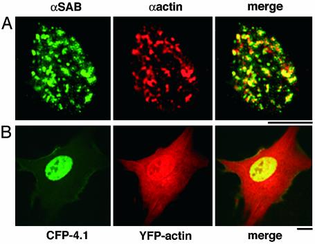
Fig. 1.
Colocalization of nuclear actin and 4.1 in human diploid fibroblasts. (A) Double-label immunofluorescence of 4.1 SABD epitopes (green) and actin (red) in WI38 cells extracted in situ to prepare nuclear matrix. The merged image shows a high degree of coincidence (yellow). (B) Transient cotransfection of WI38 cells with YFP-actin (red) and CFP-80kD4.1R (green). Cells were imaged 4–72 h posttransfection. The 24-h image presented shows expression of YFP-actin in both cytoplasm and nucleus, whereas CFP-4.1 is exclusively nuclear, generating coincident nuclear signals (yellow). (Bar, 10 μm.)

