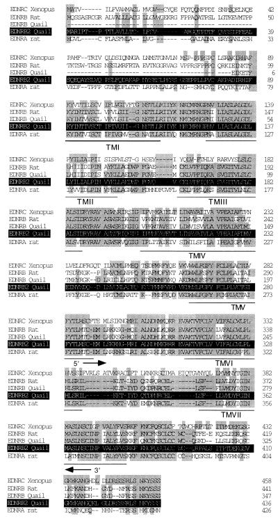
Figure 1.
Amino acid alignment of the quail EDNRB2 (in white on a black background), quail EDNRB (16), rat EDNRB (18), Xenopus EDNRC (14), and rat EDNRA (43). The residues in common between EDNRB2 and other endothelin receptor are presented on a gray background. Transmembrane domains are underlined. The region of the molecule whose sequence has been amplified by RT-PCR is located between the two arrows (representing the oligonucleotides).

