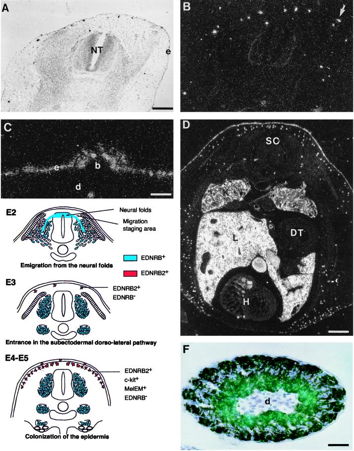
Figure 3.
EDNRB2 expression pattern in the quail embryo. (A and B) In situ hybridization with EDNRB2 probe on a transverse section of E4 quail embryos (stage 18Z) showing labeled cells, presumably melanoblasts, migrating under the ectoderm (e). Some of them have already entered the ectoderm (arrow). NT, neural tube. (C) Detail of a E6 quail feather bud. Individual cells in the epidermis (e) and in the deep dermis (d) express strongly EDNRB2. These cells are especially numerous in the dorsal feather buds (b). (D) In situ hybridization with EDNRB2 probe on transverse section of E6 quail embryos (stage 23Z). General view showing a strong hybridization signal in the liver (L), kidney (K), and skin. The white patches in the heart are an artifact due to the blood, visualized in dark field at low magnification. SC, spinal cord; DT, digestive tract; H, heart. (E) Schematic representation of the respective expression pattern of the two EDNRB genes during avian NC cell development. During emigration from the neural tube, the NC cells express EDNRB, whereas as soon as they become engaged in the dorsolateral pathway, they start expressing EDNRB2 and stop expressing EDNRB. (F) EDNRB2 expression in pigmented melanocytes, as shown by in situ hybridization with EDNRB2 probe on transverse section of E14 quail feather filament. The labeling is concentrated at the basis of the barb ridges near the dermal pulp of the feather filament (d) where the cell bodies of the melanocytes are located. The melanocytes extend processes toward the external keratinocytes and transfer their melanosomes, which form clumps of pigment at the periphery of the feather filament. Bright field (A); dark field (B–D); and epipolarized illumination (F). [Bars = 122 μm (A and B), 61 μm (C), 183 μm (D), and 64 μm (F).]

