Fig. 1.
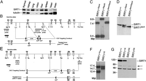
Generation of SIRT1-deficient mice. (A) Western analysis of SIRT1 and tubulin expression in WT adult mouse tissues. (B) A schematic diagram of the germ-line SIRT1 locus, KOII targeting vector, and SIRT1Δex4 allele. SIRT1 exons comprising the conserved catalytic domain are dark gray. The relative locations of the 5′, 3′, and internal KOII probes are indicated. Restriction sites: BgI, BglI; N, NotI; S, SalI; H, HindIII; E, EcoRI; Bg, BglII; B, BamHI; Pac, PacI; Swa, SwaI. (C) Southern blot of SIRT1+/Δex4, SIRT1+/NeoR, and SIRT1Δex4/Δex4 BglII-digested DNA with the internal KOII probe. (D) Western blot of SIRT1+/+, SIRT1+/Δex4, and SIRT1Δex4/Δex4 MEFs. SIRT1 and SIRT1Δex4 proteins are indicated. (E) A schematic diagram of the germ-line SIRT1 locus, SKO targeting vector, and SIRT1Δ allele, which are depicted as in A. (F) Southern blot of SIRT1+/Δneo and SIRT1+/Δ BglI/SwaI-digested DNA with the 5′SKO probe. (G) Western blot of SIRT1+/+, SIRT1+/Δ, and SIRT1Δ/Δ MEFs with anti-SIRT1 antibodies. Arrows indicate crossreacting bands that control for total protein.

