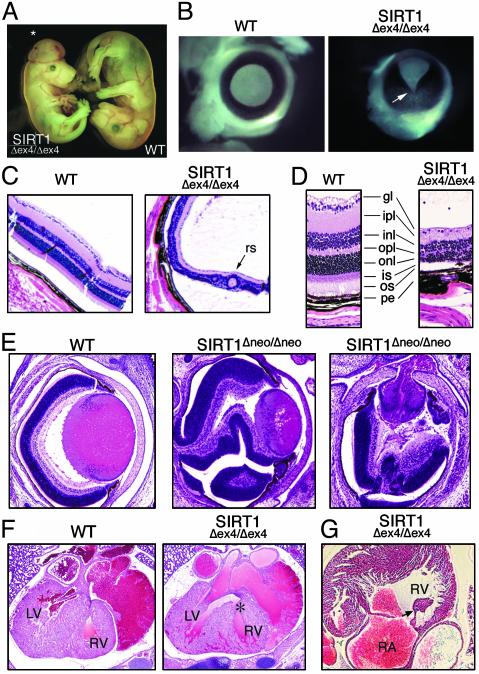
Fig. 2.
Developmental abnormalities in SIRT1-deficient mice. (A) WT and SIRT1Δex4/Δex4 E16.5 embryos. *, exencephaly in the SIRT1Δex4/Δex4 embryo. (B) Whole-mount eyes from E16.5 embryos. Arrow indicates open optic fissure in the SIRT1Δex4/Δex4 eye. (C) Retinal sections from 5-week-old mice. rs, rosette-like structure. (D) An enlargement of C. gl, ganglion cell layer; ipl, inner plexiform layer; inl, inner nuclear layer; opl, outer plexiform layer; onl, outer nuclear layer; is, inner segment; os, outer segment; pe, pigmented epithelium. (E) Retinal sections from E18.5 WT and SIRT1Δneo/Δneo embryos. (F) Sections through E18.5 hearts. *, the VSD. LV, left ventricle; RV, right ventricle. (G) Section through E18.5 SIRT1Δex4/Δex4 heart showing elongated atrioventricular valve (arrow).

