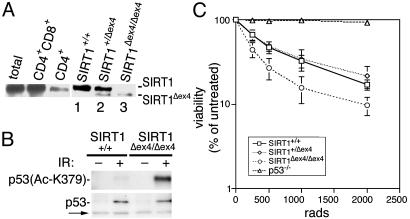
Fig. 4.
Increased thymocyte apoptosis in SIRT1-deficient cells. (A) Western analysis of SIRT1 and SIRT1Δex4 protein in thymocytes of the indicated genotypes. (B) Western analysis of acetylated p53 in p53 IPs from SIRT1+/+ and SIRT1Δex4/Δex4 thymocytes, at 2.5 h after mock (–) or 500 rads IR (+), in the presence of 1 μM TSA. Western analyses on the IP inputs show total p53 levels, and the arrow indicates a crossreacting band that controls for total protein. (C) The percent nonapoptotic cells in SIRT1+/+, SIRT1+/Δex4, SIRT1Δex4/Δex4, and p53–/– CD4+ thymocytes after IR is shown.

