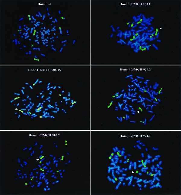
Figure 3.

FISH analysis of parental HONE1 cells and HONE1–chromosome 3 microcell hybrids. The copies of chromosome 3 are visualized by in situ hybridization using a chromosome 3-specific library probe. The transferred chromosome 3 (via microcell fusion) is identified in each case by an arrowhead. The identity of the transferred chromosome is indicated by identifying the A9–chromosome 3 donor cell.
