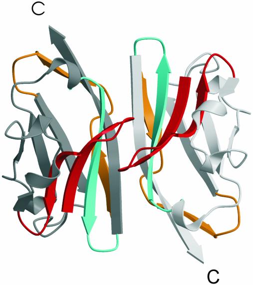
Fig. 4.
Schematic representation of the crystallographic head-to-tail dimer of MOGED. Mapping of the encephalitogenic peptides onto the dimer: orange, residues 1–22; red, residues 35–55; cyan, residues 92–106. All map onto the face of the β-sheet that mediates dimer contacts. Protomers A and B are colored light gray and dark gray, respectively.

