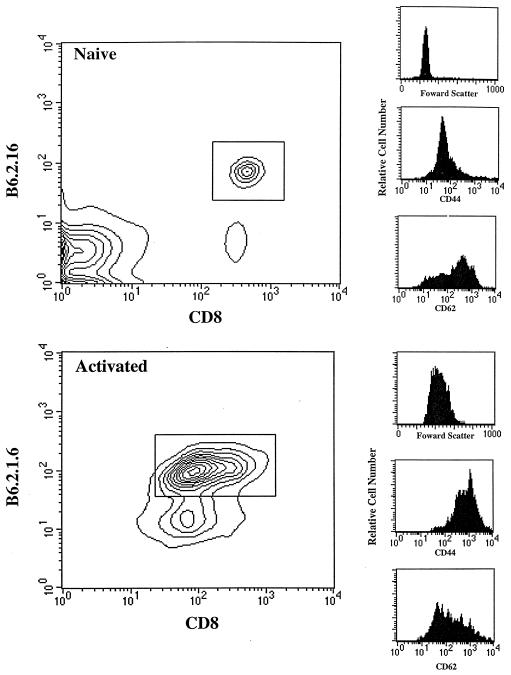
Figure 1.
Activation of female B6.2.16 T cells by H-Y peptide in vitro. Pooled spleen and mesenteric lymph node cells (5 × 10⁵) from B6.2.16 transgenic mice were stained with the following mAbs: T3.70-FITC [anti-B6.2.16 clonotype (18)], anti-CD8-APC, and either anti-CD44-PE or anti-CD62-PE, and then analyzed by FACS. The expression level of CD44 and CD62 was determined on gated B6.2.16 CD8+ cells. Activated B6.2.16 CD8+ cells were generated by incubating naive cells (106/ml) with the H-Y peptide (10−7 M) for 3–4 days.

