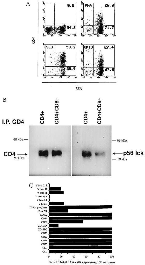
Figure 1.

(A) CD4 antigen induction in activated mature CD8+ T cells. Resting, or 6-day-old PHA-P (1 μg/ml), SEB (1 μg/ml), or anti-CD3 (200 ng/ml) activated cells were stained for both CD4 and CD8 antigen expression. Control aliquots were stained with FITC- or PE-conjugated irrelevant mouse IgG1. (B) Association of p56lck to CD4 in activated mature CD8+ T cells. PBMC depleted CD4+ T cells were activated with SEB for 6 days. CD8+ T cells (25 × 106), of which 24% were expressing CD4, were lysed and CD4 immunoprecipitated. Following SDS/PAGE electrophoresis, proteins were transferred to nitrocellulose and probed for both CD4 and p56lck. SEB (107) activated CD4+ T cells were used as positive controls. (C) Phenotypic analysis of CD8+ T cells coexpressing CD4. Seven-day-old SEB activated CD4-depleted PBMC (0% CD8−CD4+) were simultaneously stained with PE-conjugated anti-CD4 and various FITC-conjugated mAb. The percentage of cells expressing a specific marker was determined after acquisition and analysis of 10,000 events.
