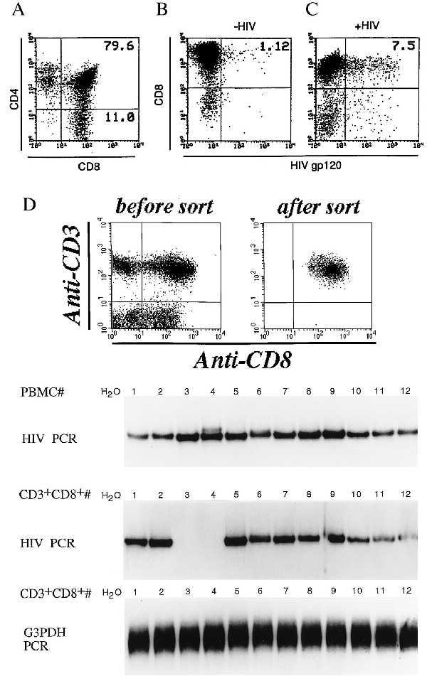
Figure 4.

(A) Analysis of DP cells in activated PBMC cultures from HIV-infected individuals. PBMC from HIV-infected patients were isolated and stimulated with PHA. On days 12–19, cells were stained and analyzed for CD4 and CD8 coexpression. (B) Infection of CD8+ T cells by endogenous HIV. On days 12–19 poststimulation cells were stained for both CD8 and HIV gp120 expression and analyzed by flow cytometry. Results from one donor, representative of five, is presented. (C) Infection of CD8+ T cells from HIV-infected individuals with HIV-1 IIIB. PBMC from HIV-infected individuals were stimulated for 12–19 days with PHA and then treated with HIV-1 IIIB (multiplicity of infection, 0.1). After five days, cells were stained for surface expression of both CD8 and HIV-1 gp120 and analyzed by flow cytometry. (D) HIV DNA detection in PBMC and sorted CD3+CD8+ T cells from HIV-infected individuals. DNA from PBMC or CD3+CD8+ sorted T cell population from 12 HIV-infected individuals was isolated and subjected to PCR for the presence of HIV DNA sequences. G3PDH gene was amplified as positive control and water as negative control. (Top) The dot plot profile of one donor, representative of 12, before and after sorting of CD3+CD8+ T cells.
