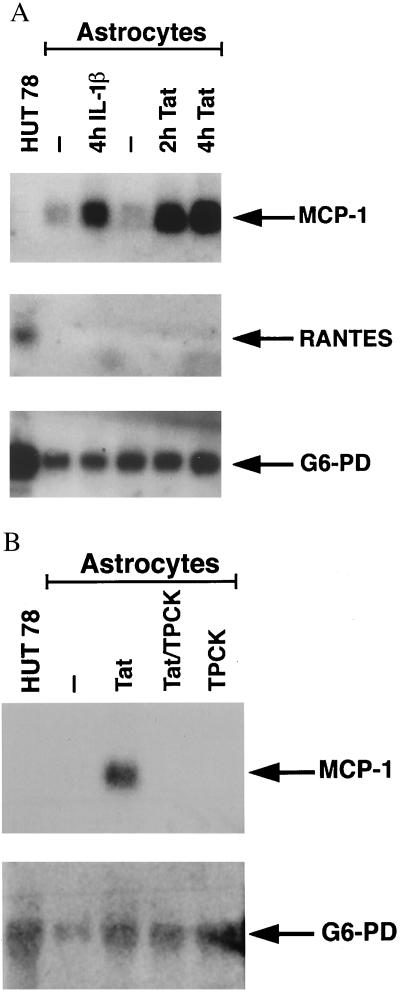
Figure 1.
(A) Total RNA was extracted from T lymphoblastoid HUT 78 cells (ATCC) and from variously treated human astrocytes. Ten micrograms per lane were then run on a 1% agarose–6% formaldehyde gel. After transfer of RNA to nitrocellulose, the blot was probed with the full-length cDNA for MCP-1 and, subsequently, RANTES. RNA from untreated astrocytes was run in lanes 2 and 4. Both 4 hr stimulation with 5 ng/ml of interleukin-1β (lane 3) and 2 or 4 hr stimulation with 100 nM of Tat (lanes 5 and 6) were associated with an increase in astrocytic MCP-1 expression. Astrocytic expression of RANTES was not detected. (B) Similar experiment except that RNA from HUT 78 cells (lane 1) was compared with RNA from astrocytes that were unstimulated (lane 2) or stimulated for 2 hr with 100 nM of Tat (lane 3), 25 μM of TPCK followed by 100nM of Tat (lane 4), or 25 μM of TPCK (lane 5). In A and B, the G6-PD probe was used as a control (24).

